
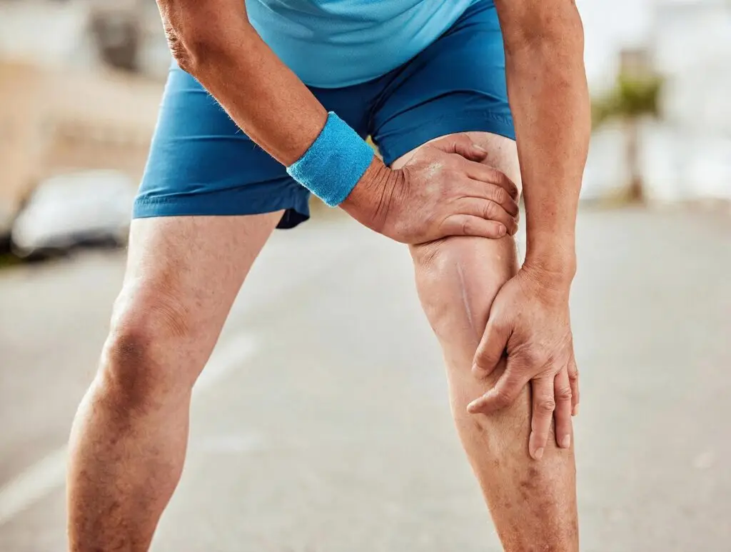
Knee pain can develop for a range of reasons, from gradual wear and tear through to injury, instability, inflammation or underlying joint conditions. Depending on the cause, knee symptoms may include pain, swelling, stiffness, catching, locking, weakness or a feeling that the knee may give way. These issues can affect your ability to walk comfortably, use stairs, exercise, work, kneel, squat or participate in everyday activities.
If your symptoms are persistent, worsening or limiting your movement, a thorough assessment can help identify the underlying cause and guide appropriate treatment options based on your individual needs, activity level and overall joint health.
Knee pain is a common symptom that can affect people of all ages and may develop gradually or occur suddenly. It can be felt at the front, inside, outside or back of the knee, depending on the underlying cause.
In some cases, knee pain is related to changes within the joint itself, such as cartilage wear, arthritis or inflammation. It may also arise from surrounding structures, including the ligaments, tendons, muscles, meniscus or bursae.
Common causes of knee pain may include:
The pattern, location and severity of your pain can provide important clues about the underlying cause. If your symptoms are ongoing, worsening or impacting your daily activities, further assessment may help determine the cause and guide appropriate management.
Treatment options will depend on the underlying condition and may range from physiotherapy, activity modification, bracing or injections through to surgical options in selected cases.
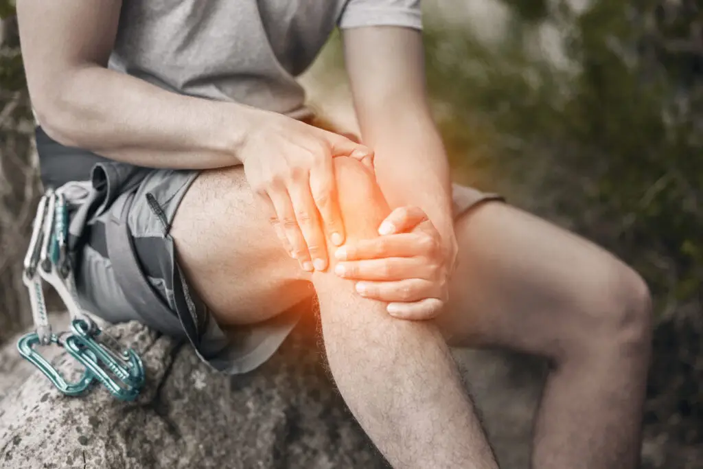
Knee arthritis, most commonly osteoarthritis, is a condition where the smooth cartilage lining the knee joint gradually wears down over time. This can lead to increased friction within the joint, resulting in pain, stiffness, swelling and reduced movement.
Osteoarthritis of the knee typically develops slowly and is more common with increasing age, although it can also occur earlier due to previous injury, meniscal damage, ligament injury, altered knee alignment or increased joint loading over time.
Common symptoms may include:
As the condition progresses, symptoms may become more persistent and begin to impact daily activities, mobility and quality of life.
Treatment options for knee arthritis:
Management depends on the severity of symptoms, the degree of joint damage and how much the condition affects your function. Early treatment may include activity modification, physiotherapy, weight management where appropriate, pain management strategies, bracing or injections. These options may help manage symptoms, although they do not reverse the underlying cartilage wear.
When symptoms become more advanced and non-surgical treatments are no longer providing enough relief, surgical treatment may be considered. The most commonly performed procedure for advanced knee arthritis is total knee replacement, which aims to relieve pain and improve function by replacing the damaged joint surfaces.
In selected cases, different surgical options may be discussed, including:
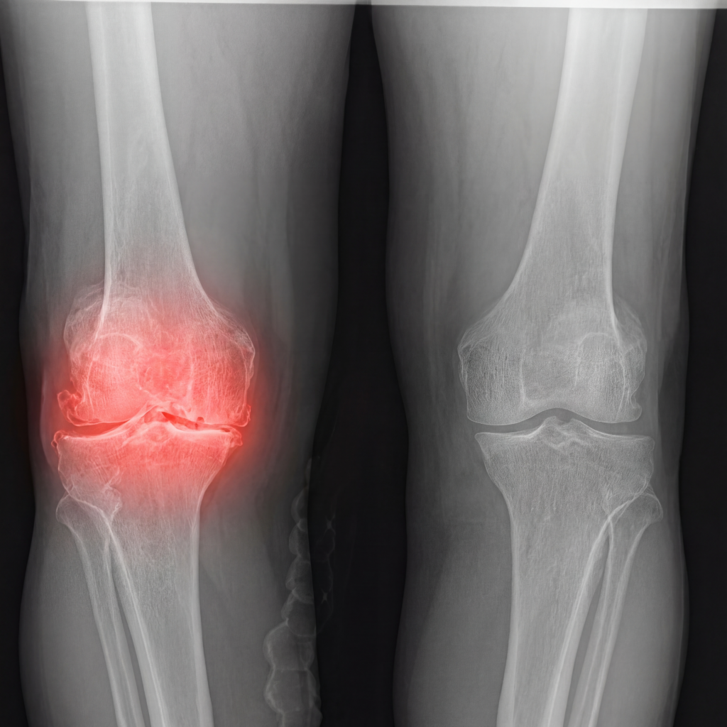
A thorough assessment can help confirm the diagnosis, identify which part of the knee is affected and guide the most appropriate treatment plan based on your individual needs.
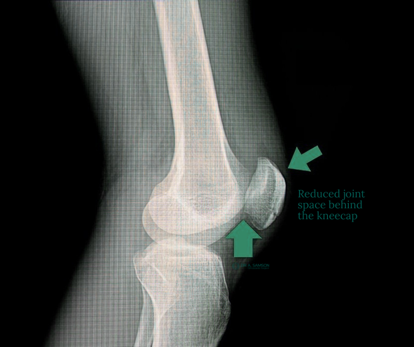
Patellofemoral arthritis is a form of knee arthritis that affects the joint between the kneecap, known as the patella, and the groove at the front of the thigh bone, known as the trochlea. This part of the knee helps the kneecap glide smoothly as the knee bends and straightens.
When the smooth cartilage in the patellofemoral joint gradually wears down, it can lead to pain at the front of the knee, stiffness, swelling and grinding sensations. Symptoms are often more noticeable during activities that place extra load on the kneecap, such as stairs, squatting, kneeling, rising from a chair or walking on hills.
Patellofemoral arthritis may develop as part of general knee osteoarthritis, or it may occur due to previous kneecap instability, patella dislocation, trauma, altered knee alignment, trochlear dysplasia or increased joint loading over time.
Common symptoms may include:
Management depends on the severity of symptoms, the degree of cartilage wear and whether arthritis is limited to the patellofemoral joint or also affects other areas of the knee. Early treatment may include activity modification, physiotherapy, strengthening exercises, weight management where appropriate, pain management strategies, bracing or injections. Physiotherapy often focuses on improving quadriceps, hip and gluteal strength, as this may help support kneecap tracking and reduce load through the front of the knee.
When symptoms become more advanced and non-surgical treatments are no longer providing enough relief, surgical treatment may be considered. The most appropriate procedure depends on the pattern of arthritis, knee alignment, kneecap tracking and overall joint health.
In selected cases, surgical options may include:
A thorough assessment can help confirm the diagnosis, identify the extent of joint involvement and guide the most appropriate treatment plan based on your symptoms, imaging findings and individual needs.
Sports knee injuries can occur during running, jumping, landing, twisting, pivoting or sudden changes in direction. These injuries are common in activities such as football, netball, basketball, soccer, skiing, running and court sports, where the knee is placed under repeated load or exposed to sudden force.
A sports-related knee injury may affect the ligaments, meniscus, cartilage, tendons, muscles or kneecap joint. Some injuries occur suddenly after a fall, collision or twisting movement, while others develop gradually due to overuse, repetitive loading or poor movement control.
Common sports knee injuries may include:
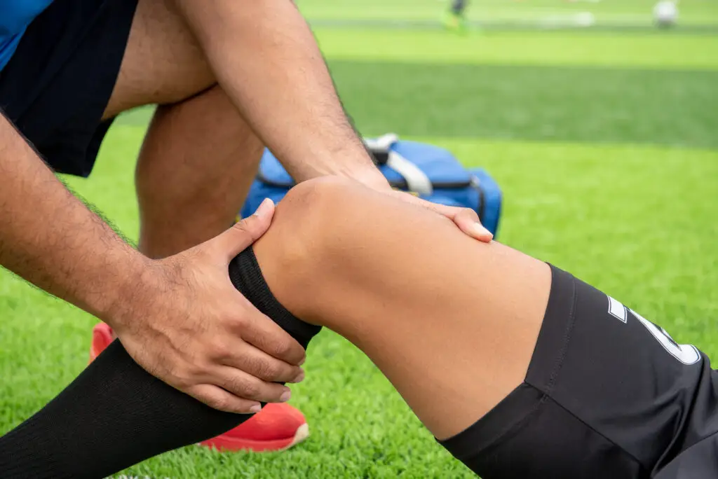
Symptoms can vary depending on the type and severity of injury, but may include:
Some sports knee injuries settle with non-surgical management, while others may require further investigation and treatment, particularly if there is ongoing swelling, instability, locking, loss of movement or difficulty returning to sport.
Management depends on the type of injury, the structures involved, your symptoms, activity level and goals for returning to sport or daily function. Early treatment may include rest, ice, activity modification, bracing, physiotherapy and pain management strategies. Further assessment may include clinical examination and imaging, such as X-rays or MRI, to help identify the injured structures and guide appropriate treatment. In selected cases, surgical treatment may be considered, particularly for injuries involving significant ligament damage, meniscal tears, cartilage injuries or recurrent kneecap instability.
Depending on the injury, surgical options may include:
Anterior cruciate ligament, or ACL, tears are common knee injuries that often occur during sport or activities involving sudden stopping, landing, pivoting, twisting or changing direction. The ACL is one of the main ligaments inside the knee and helps control stability, rotation and forward movement of the shin bone in relation to the thigh bone.
An ACL tear may occur during contact or non-contact injuries. In many cases, the injury happens when the knee twists awkwardly, buckles on landing, or changes direction suddenly while the foot remains planted. ACL injuries are commonly seen in sports such as football, netball, basketball, soccer, skiing and court sports.
An ACL tear may occur on its own or in combination with other knee injuries, such as meniscal tears, cartilage damage, bone bruising or collateral ligament injuries.
Common symptoms of an ACL tear may include:
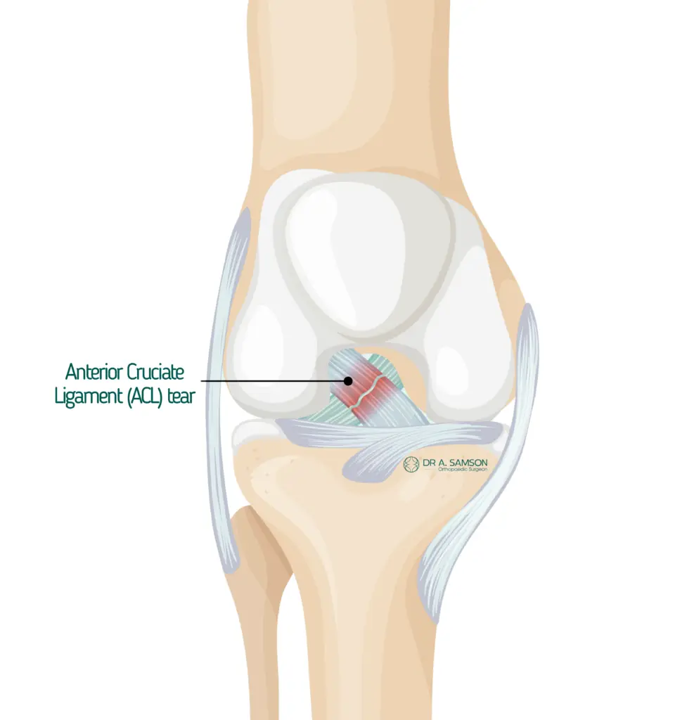
Some people are able to walk after an ACL injury, while others find it difficult to put weight through the knee. Symptoms can vary depending on the severity of the tear and whether other structures within the knee have also been injured.
Diagnosis of ACL tears
A thorough assessment can help determine whether the ACL has been injured and whether there are associated injuries within the knee.
Assessment may include a discussion about how the injury occurred, your symptoms, your sporting or activity goals, and a clinical examination to assess knee swelling, movement, ligament stability and areas of tenderness. Imaging may also be recommended. X-rays can help assess for fractures or bony injury, while an MRI scan can provide more detailed information about the ACL, meniscus, cartilage and other soft tissues within the knee.
Treatment options for ACL tears
Treatment for an ACL tear depends on the severity of the injury, whether other structures have been damaged, your symptoms, knee stability, activity level, age, lifestyle and goals for returning to sport or daily function. In some cases, non-surgical management may be appropriate. This may involve physiotherapy, strengthening, activity modification and bracing where required. Non-surgical care may be considered for people who do not experience ongoing instability and who do not need to return to pivoting or high-demand sports. For people who have ongoing instability, recurrent giving way, associated meniscal or cartilage injuries, or a goal to return to pivoting sports, ACL reconstruction surgery may be considered.
ACL reconstruction involves replacing the torn ligament with a graft to help restore stability to the knee. The graft may be taken from your own tissue, such as the hamstring tendon, patellar tendon or quadriceps tendon, depending on your individual needs and the surgeon’s recommendation.
The procedure is commonly performed using arthroscopic, or keyhole, surgical techniques. Small incisions are made around the knee, and a camera is used to view the inside of the joint. The torn ACL is removed, tunnels are created in the thigh bone and shin bone, and the graft is positioned to recreate the function of the ACL.
If there are associated injuries, such as a meniscal tear or cartilage damage, these may also be treated during the same operation where appropriate.
Meniscus tears are common knee injuries that affect the C-shaped cartilage pads inside the knee joint. Each knee has two menisci, the medial meniscus on the inside of the knee and the lateral meniscus on the outside. These structures help absorb shock, distribute load, improve joint stability and protect the cartilage surfaces of the knee. A meniscus tear may occur suddenly during sport or activity, particularly with twisting, pivoting, squatting or changing direction. Tears can also develop gradually over time due to age-related wear, arthritis or repeated loading of the knee.
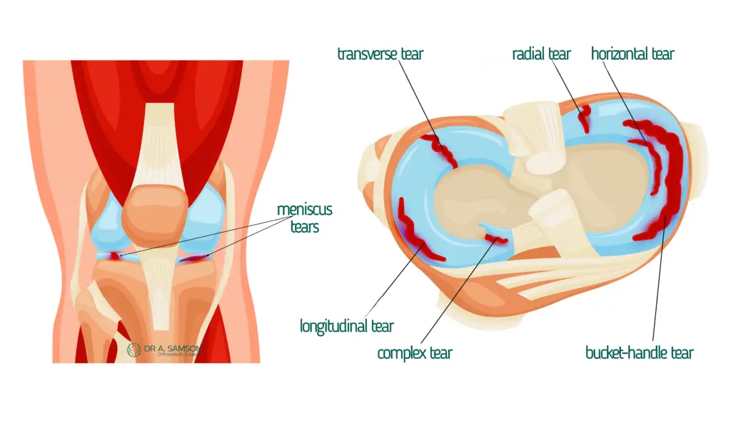
Symptoms can vary depending on the size, location and type of tear, as well as whether there is underlying arthritis or cartilage wear within the knee.
Treatment options for meniscus tears
Management depends on the type of meniscus tear, your symptoms, activity level, age, knee alignment and the condition of the surrounding joint surfaces. Further assessment may include a clinical examination and imaging, such as X-rays or MRI, to help confirm the diagnosis and identify any associated knee conditions.
Some meniscus tears may settle with non-surgical management, including physiotherapy, activity modification, pain management strategies and, in some cases, injections. These options may help reduce symptoms and improve function, particularly when the tear is related to degenerative change or arthritis.
In selected cases, surgical treatment may be considered, especially if there is ongoing pain, locking, catching, swelling or difficulty returning to activity. Surgical options may include meniscus repair, where the torn tissue is stitched where healing is possible, or partial meniscectomy, where the damaged portion of the meniscus is removed while preserving as much healthy tissue as possible.
A thorough assessment can help determine the type of meniscus tear, whether other structures in the knee are involved, and guide the most appropriate treatment plan based on your symptoms, lifestyle and individual needs.
Knee ligament injuries occur when one or more of the ligaments that help stabilise the knee are stretched, partially torn or completely torn. The main knee ligaments include the anterior cruciate ligament, or ACL, posterior cruciate ligament, or PCL, medial collateral ligament, or MCL, and lateral collateral ligament, or LCL. These ligaments work together to control knee stability, alignment and movement, particularly during twisting, pivoting, landing, side-to-side movement and changes in direction.
A knee ligament injury may occur during sport, a fall, a collision, a twisting injury or direct trauma to the knee. These injuries are commonly seen in activities such as football, netball, basketball, soccer, skiing and court sports, although they can also occur during everyday accidents.
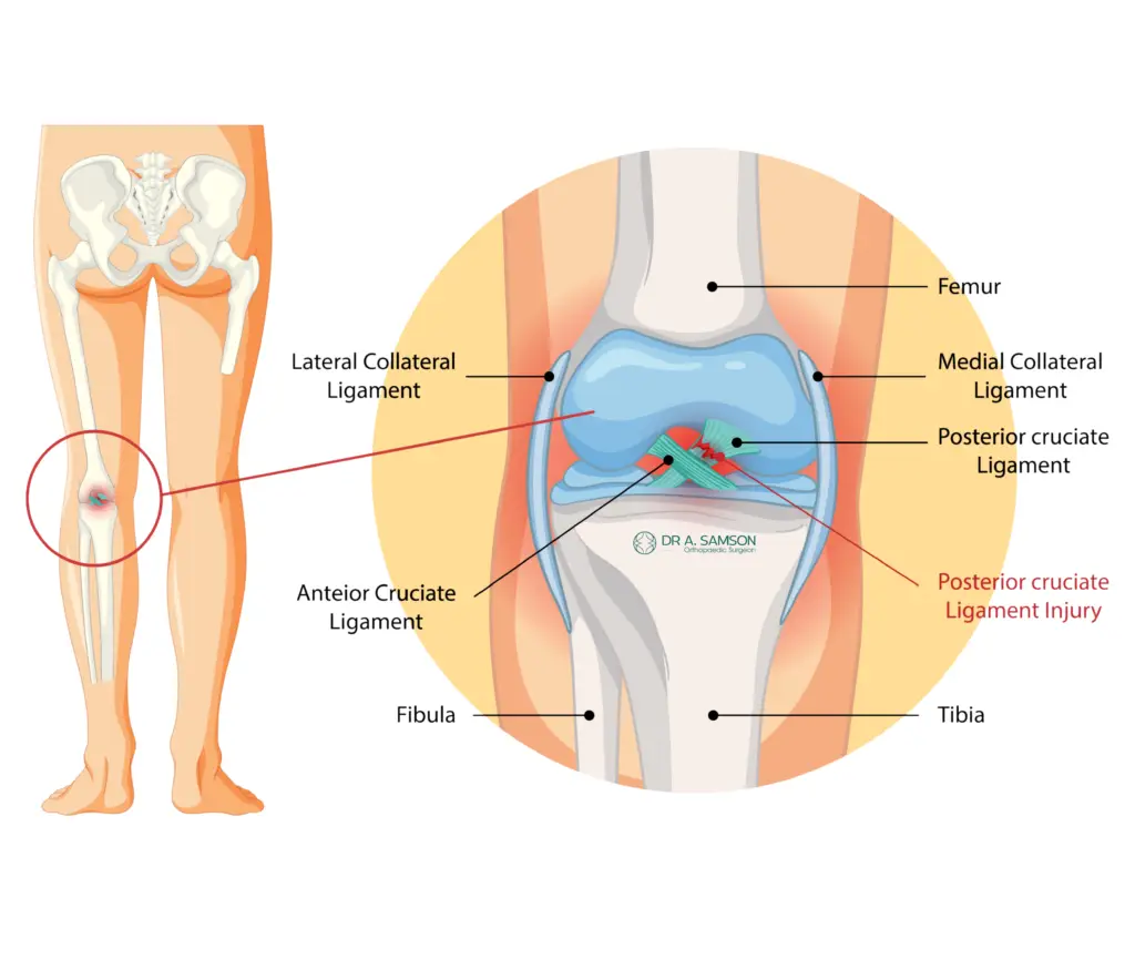
Symptoms can vary depending on which ligament is injured, whether the tear is partial or complete, and whether other structures in the knee are also affected, such as the meniscus, cartilage or bone.
Treatment options for knee ligament injuries
Management depends on the ligament involved, the severity of the injury, your symptoms, knee stability, activity level and goals for returning to sport or daily function. Further assessment may include a clinical examination and imaging, such as X-rays or MRI, to help identify the injured ligament and assess for associated knee injuries.
Some ligament injuries may settle with non-surgical management, including rest, bracing, physiotherapy, strengthening, activity modification and gradual return to activity. This may be appropriate for selected partial ligament injuries or lower-grade sprains where the knee remains stable.
In selected cases, surgical treatment may be considered, particularly when there is significant instability, a complete ligament rupture, multiple ligament injuries, or difficulty returning to sport or normal function. Surgical options may include ligament repair or ligament reconstruction, depending on the type and severity of injury. A thorough assessment can help identify which ligament has been injured, determine the severity of the damage, and guide a treatment plan based on your knee stability, lifestyle, sporting goals and individual needs.
Knee cartilage damage, also known as an osteochondral defect, when the underlying bone is involved, refers to injury or wear affecting the smooth cartilage surface that lines the ends of the bones inside the knee joint. This cartilage allows the joint surfaces to move smoothly and helps absorb load during walking, running, squatting and other weight-bearing activities. Cartilage damage may occur suddenly after an injury, such as a fall, twisting movement or direct impact to the knee. It may also develop gradually over time due to repetitive loading, previous knee injury, altered knee alignment, arthritis or joint degeneration.
An osteochondral defect may affect the cartilage alone or involve both the cartilage and the underlying bone. These injuries can occur in different parts of the knee, including the thigh bone, shin bone or kneecap joint.
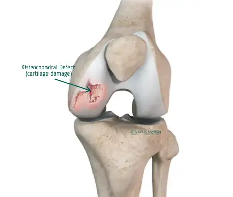
Symptoms can vary depending on the size, location and depth of the cartilage injury, whether the underlying bone is involved, and whether there are other knee conditions such as meniscal tears, ligament injuries or arthritis.
Treatment options for knee cartilage damage
Management depends on the type and severity of cartilage damage, the area of the knee involved, your symptoms, activity level, knee alignment and whether there is underlying arthritis or joint degeneration. Further assessment may include a clinical examination and imaging, such as X-rays or MRI, to help identify the location and extent of the cartilage injury and assess the surrounding joint surfaces.
Some cartilage injuries may be managed with non-surgical treatment, including physiotherapy, activity modification, strengthening, weight management where appropriate, pain management strategies or injections. These options may help manage symptoms, although they do not restore the damaged cartilage surface. In selected cases, surgical treatment may be considered, particularly when symptoms are ongoing, the defect is localised, or there are mechanical symptoms such as catching or locking. Surgical options may include arthroscopic knee surgery, cartilage procedures, or treatment of associated injuries such as meniscal or ligament damage. A thorough assessment can help determine the type of cartilage injury, whether the underlying bone is involved, and guide the most appropriate treatment plan based on your symptoms, knee function, activity goals and individual needs.
A failed knee replacement refers to a knee replacement that is no longer functioning as expected or is causing ongoing pain, stiffness, instability or reduced mobility. While many knee replacements function well for many years, some may require further assessment if symptoms develop or the implant becomes worn, loose, unstable or affected by infection. Symptoms may occur soon after surgery or develop gradually over time. A failed knee replacement may be related to implant loosening, wear of the implant surfaces, infection, instability, stiffness, malalignment, fracture, soft tissue imbalance or ongoing pain around the knee.
Symptoms can vary depending on the underlying cause. Some people experience gradual discomfort and reduced function, while others may develop sudden pain after a fall, injury or infection.
Treatment options for a failed knee replacement
Management depends on the cause of symptoms, the condition of the implant, the surrounding bone and soft tissues, and how much the knee is affecting your daily function. Further assessment may include a clinical examination, review of your previous surgery and implant details, blood tests, X-rays and, in some cases, additional imaging such as CT or bone scans. These investigations help identify whether the implant is loose, worn, malaligned, infected or affected by another issue. Some symptoms may be managed without further surgery, depending on the cause. This may include physiotherapy, pain management strategies, activity modification or monitoring over time.
In selected cases, revision knee replacement surgery may be considered. This involves removing part or all of the existing knee replacement and replacing it with new components. Revision surgery may be more complex than the original knee replacement, particularly if there is bone loss, infection, ligament imbalance or significant stiffness. A thorough assessment can help identify the cause of pain or reduced function after knee replacement and guide the most appropriate treatment plan based on your implant, symptoms, mobility and individual needs.
Dr Anthony Samson welcomes new patients with a valid referral from a General Practitioner (GP) or Specialist.