

Every hip joint is different. The size, shape and alignment of your hip can vary depending on your natural anatomy, the severity of arthritis, previous injury, bone quality and the way your hip has changed over time.
Patient-specific planning allows these factors to be assessed in detail before surgery. Dr Samson combines this planning with the anterior approach to total hip replacement, allowing implant selection and positioning to be tailored to your hip anatomy, alignment and individual requirements.
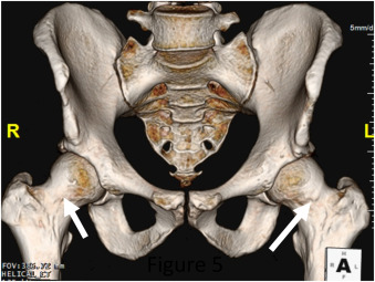
As part of this process, you will need to have a CT scan of your hip before surgery. Dr Samson’s team will arrange this imaging for you.
The scan provides detailed information about the three-dimensional shape and structure of your hip joint and pelvis and forms the basis of your patient-specific surgical plan.
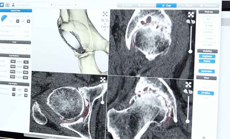
The CT images are then used to create a 3D digital model of your hip anatomy. This model helps guide detailed pre-operative planning and allows Dr Samson to assess your hip joint in relation to your pelvis, leg alignment and overall biomechanics.
Dr Samson reviews your imaging, including X-rays and CT imaging, to develop a surgical plan tailored to your individual anatomy, hip condition and surgical needs.
Advanced digital planning software is used to support this process and assists with the assessment of your:

Once the surgical plan has been developed, Dr Samson uses the digital planning software and 3D model to define the planned implant size and position for your hip replacement. In collaboration with engineers, a patient-specific surgical guide is then created based on your 3D model and surgical plan. The guide is designed specifically to match your anatomy and assist with reproducing the planned component position during surgery. The guide is sterilised and sent to the hospital before your operation.
During surgery, Dr Samson uses your patient-specific guide to help translate the pre-operative plan to your hip and assist with implant positioning.